基于Mamba改进的3D肝脏及肝肿瘤CT图像分割

打开文本图片集
中图分类号:TP391.4 文献标识码:A 文章编号:2096-4706(2026)03-0082-06
Improved 3D Liver and Liver Tumor CT Image Segmentation Based on Mamba
GUOJiahao,HUHuaifei (South-Central Minzu University,Wuhan 43oo74, China)
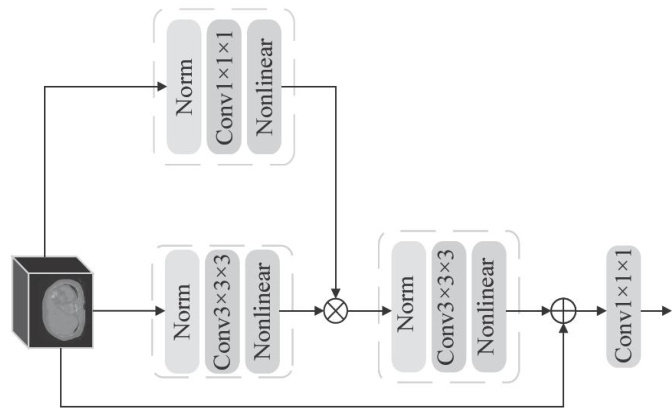
Abstract: Accurate segmentation of liver and liver tumors in three-dimensional Computed Tomography (CT) images is crucialforclinicaldiagnosisandtreatmentplanning.ToaddresstheissuesthatexistingConvolutionalNeuralNetwork(CNN) segmentationmethodsstrugle tocapturelong-rangedependenciesduetolimitedreceptivefelds,andTransformersegmentation methodsarerestricted inapplicationonthree-dimensional medicalimages with limitedsamples,this paper proposesanew 3D medicalimage segmentation model based on Mamba.The model contains a 3D gated spatial convolution module andachannel shuffle upsampling module, which extract spatial correlation features and common features,respectively.The two modules synergistically enhance themodeling abilityofthe modelfor global tructures,while simultaneouslyensuring precisebondary positioning.ExperimentalresultsontheLiTS2o17datasetshow thatthe Dice SimilarityCoeffcient (DSC)of liversegmentation reaches 96.42% ,and theDSC of liver tumor segmentation reaches 70.70% .In the generalization experiment on the 3D-IRCADb dataset,the DSCsof liver and liver tumor segmentationreach 96.79% and 67.10% ,respectively.Resultsof multiple comparative experiments further verify the superiority and robustness of the proposed model in segmentation performance.
Keywords: liver tumor segmentation; Mamba; State Space Model; Deep Learning
0 引言
医学图像分割在疾病诊断和治疗规划中发挥着重要作用,这一特点在肝肿瘤治疗领域尤为显著[1-3]精准的分割结果将直接影响手术方案的设计与术后评估。(剩余11732字)